Core Platforms & Infrastructure
- Research
- Core Platforms & Infrastructure
State-of-the-art facilities equipped with new technologies & infrastructure
Members of the MNN operate and utilize five core facilities vital to the continued success of ongoing research projects and our institutions. These core platforms increase our international competitiveness, enhances the ability to recruit and retain faculty and trainees, and elevates neuroscience research in Manitoba.
Learn more about our platforms below.
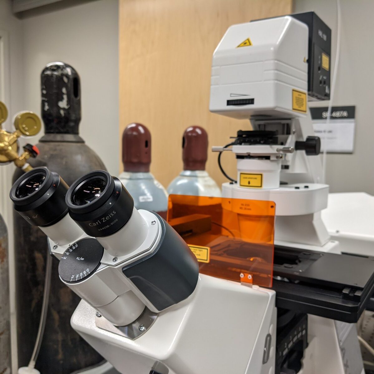
The LCIF provides access to advanced microscopy systems for imaging both live and fixed cells at low cost for researchers in Manitoba. There are currently 4 microscopy systems available, including a confocal microscope, two 2-photon systems, and a laser microdissection microscope. The LCIF is maintained and operated by the Neuroscience Research Program under the supervision of Dr. Michael F. Jackson. Three systems are located on-site on SR4 of the Kleysen Institute for Advanced Medicine, while the intravital 2-photon is located in BMSB Central Animal Care.
CHRIM
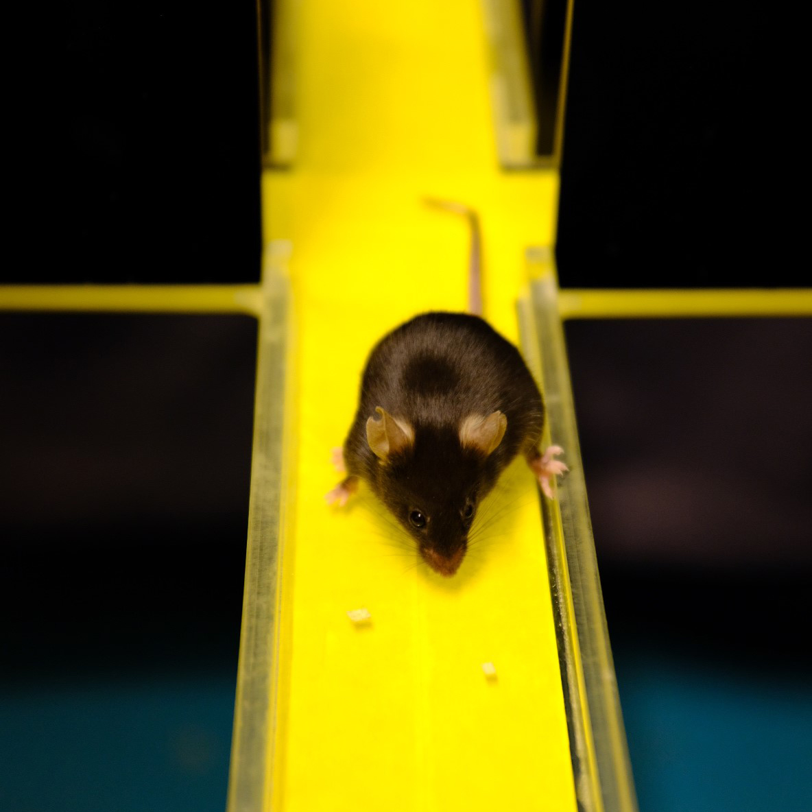
Animal behavioral core locating in Children’s Hospital Research Institute of Manitoba (CHRIM) under Dr. Kauppinen’s guardianship has state-of-the-art EthoVision® XT video tracking system with Basler Ace monochrome and IR-sensitive camera with Gigabit Ethernet interface for automatically recording animal activity, movement and behavior in any type of arena, enclosure, or maze in day light or dark. With this system one can analyze standard parameters like velocity, distance, and time spent in a certain zone, but allow also in-depth analysis providing accurate measures for e.g. time spent immobile, animal’s position, body elongation, rotations and zone entrances.
The core has equipment to analyze following functions:
| Arena, enclosure | Motor function | Learning and memory | Anxiety/depression |
| Open field with opal walls (rat, mice) |
Open field test to access motility | Novel object recognition test to assess non-spatial object identification memory |
- |
| Open field with clear walls (rat, mice) |
Open field test to access motility | Novel location recognition to evaluate spatial learning |
Anxiety |
| Cylinders (rat, mice) | Locomotor asymmetry assessment | - | Forced swimming to assess depression |
| Morris Water Maze (rat) | - | Morris water maze and probe test to evaluate spatial learning and memory retaining |
- |
| Horizontal Ladder (rat) | Motor functions | - | - |
Dr. Kauppinen’s team including 3 research personnel can train you to use the software and advice on all above tests. They can also advise how to assess above mention functions with tests that do not require advanced imaging system for analysis. These include e.g. motor strength (wire hanging), movement disorders (pole test), fine motor coordination (tape test, pasta test), depression (tail suspension test) and anxiety/obsessive-compulsive disorder (marble test).
Contact
Dr. Tiina Kauppinen
SIDDIQUI LAB BEHAVIOURAL FACILITY - BMSB
The Siddiqui Lab behaviour facility is located in the basement of the Animal Care Facility (BSMB) under Dr. Siddiqui’s guardianship. The facility is equipped with tunable light system and state of the art cameras for video capturing and tracking. Recorded videos are analyzed by the AnyMaze software. Siddiqui Lab personnel may provide training in completing behaviour experiments and analysis on a collaborative basis. Currently, the Siddiqui Lab behaviour facility only caters to mouse experiments.
The core has equipment to analyze following functions:
| Equipment | Assessment |
| Open field test | Locomotion, speed, velocity, and working memory |
| Novel object recognition test | Object recognition memory, spatial learning and working memory |
| Elevated Plus Maze | Risk avoidance, anxiety |
| Morris Water Maze | Spatial learning and memory |
| Forced swim test | Depressive behaviour/helplessness |
| Sucrose preference test | Anhedonia/Depression |
| Pup retrieval test | Maternal behaviour |
| Eight arm radial maze | Spatial learning and memory |
| Accelerating rotarod | Gross motor skills |
| Three chamber social approach test | Social memory, social affiliation and social novelty |
| Reciprocal interaction test | Social communication |
| Pre-pulse inhibition | Sensory-motor gating |
| Fear conditioning tests | Cued and contextual learning and memory |
| Tail suspension test | Depression |
Additional tests for gross and fine motor skills, depression, and repetitive behaviour.
Contact
Dr. Tabrez Siddiqui
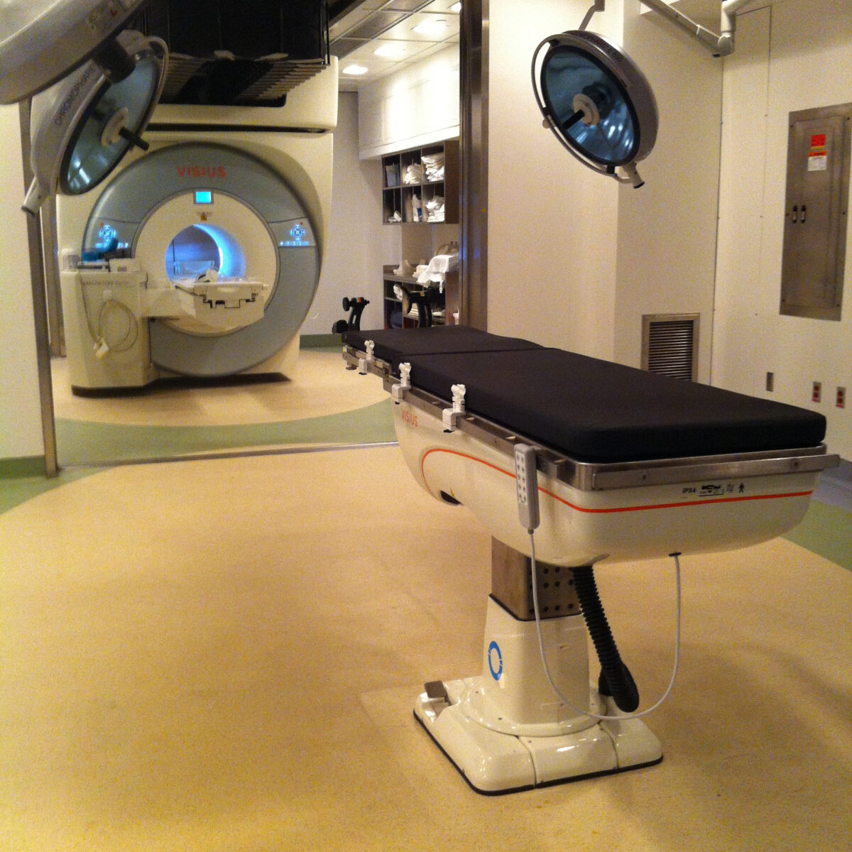
The Manitoba Neuroimaging Platform was established in 2015 with support from the Brain Canada and the HSC Foundation. It is designed to centralize innovative neuroimaging sequence and analysis development in human and animal research. The mission is to provide: standardized policies, procedures and templates for subject screening, image acquisition, and analysis; information about neuroimaging study design, protocol optimization, and data analysis through hands-on sessions and formal lectures and symposia; provide imaging expertise though imaging collaborations and support to clinical investigators; and physical resources (e.g., desk space, computers/workstations, software: MATLAB, SPM, FSL, MIPAV, ImageJ, MRIStudio, Olea, R, and SPSS) for researchers to acquire and analyze neuroimaging data. Using multimodal neuroimaging technologies, the platform is currently facilitating detailed study of brain structure and function in patients and animal models with neurodegenerative disorders including Multiple Sclerosis, Parkinson’s disease, Alzheimer’s disease and Traumatic Brain Injury. The platform provides expertise in a range of imaging techniques including MRI, PET, CT, small animal MRI, and PET-MRI. For human neuroimaging, platform components include two 3T MRI scanners (one at KIAM and other at the former Institute for Biodiagnostics, NRC). Platform components used for small animal neuroimaging include the University of Manitoba Small Animal and Materials Imaging Core Facility (SAMICF) which has capabilities in small animal CT, live animal optical imaging, 7T PET-MRI, and a two-photon microscopy system. Using these tools and physical resources, the platform provides centralized image analysis capabilities that will empower users across multiple heath research pillars to produce modelling, diagnostic and treatment deliverables they would not otherwise be able to individually achieve.
The Small Animal and Materials Imaging Core Facility (SAMICF) brings capabilities for both structural/anatomical and functional imaging to Manitoba scientists. Magnetic Resonance imaging (MRI) Positron emission tomography (PET), PET-MRI, computed tomography (CT), Single Photon Emission Computed Tomography (SPECT-CT) ultrasound and optical imaging systems are available to support academic, preclinical and industrial research. At the heart of SAMICF are state of the art MR Solutions 7T PET-MRI, Mediso Nanoscan SPECT-CT, Skyscan 1176 high resolution micro-CT, Siemens micro-PET P4, VisualSonics Vevo 2100 ultrasound and IVIS Spectrum bioluminescence/fluorescence imaging systems that can be applied to in vivo studies of small animals and materials (mineralised biomaterials, grafts etc.).
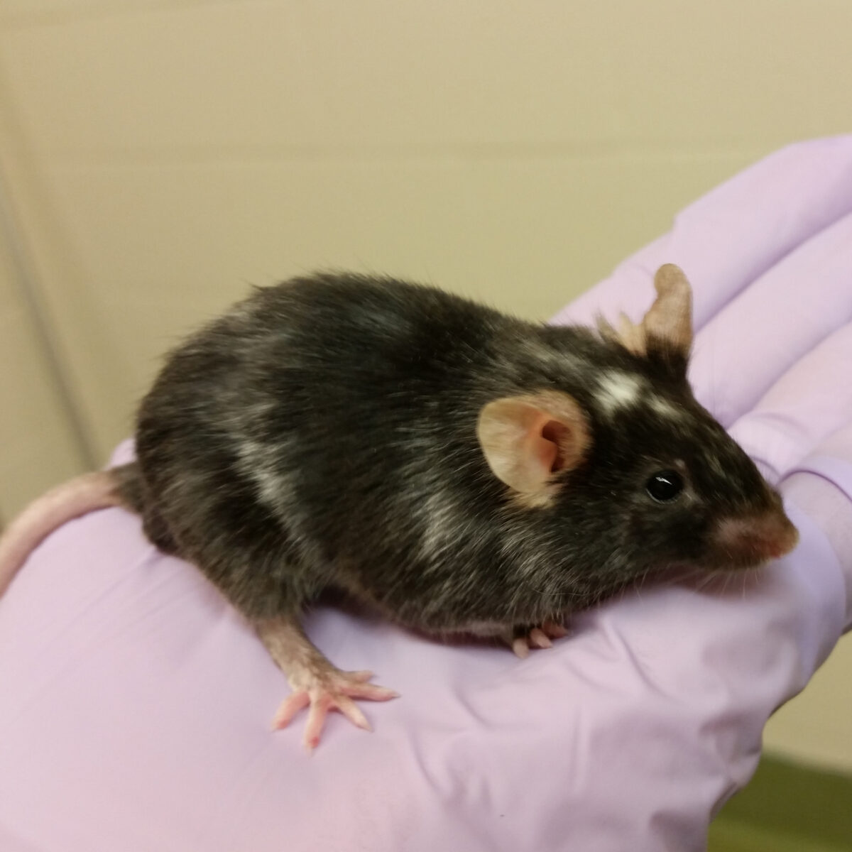
The Transgenic Facility offers a range of services for the generation, characterization and preservation of transgenic mice. Cryopreservation and re-derivation of mouse lines using sperm and embryos is available. For the generation of new animal models, we will work with individual customers to develop an approach that best suits their needs. The generation of transgenic and targeted ES cells is available and chimeric mice can be generated using either microinjection or aggregation. As well CRISPR –based approaches are now being used by the facility for both gene knockout and gene editing. Other related services include embryo collection, generation of both mouse embryonic fibroblasts and embryonic stem cells, as well as some specialized surgeries. Genotyping of mice is available. Pricing for most services is available on the web site.

The GCCRD has been designed as a high-technology facility for digital imaging and analysis. The activities have been divided into two major areas, basic research and technical services, that are envisioned to promote a better interaction between different fields of research.
The basic research component of the GCCRD is developed on collaboration with scientific groups from Canada, USA, Asia and Europe. Technical services, on the other hand, are offered on a fee basis, and these are mainly focused on fluorescent in situ hybridization, immunohistochemistry, and microdissection of biological material. The Centre does not carry out clinical diagnostics, but collaborations with clinical genetics laboratories are strongly encouraged.
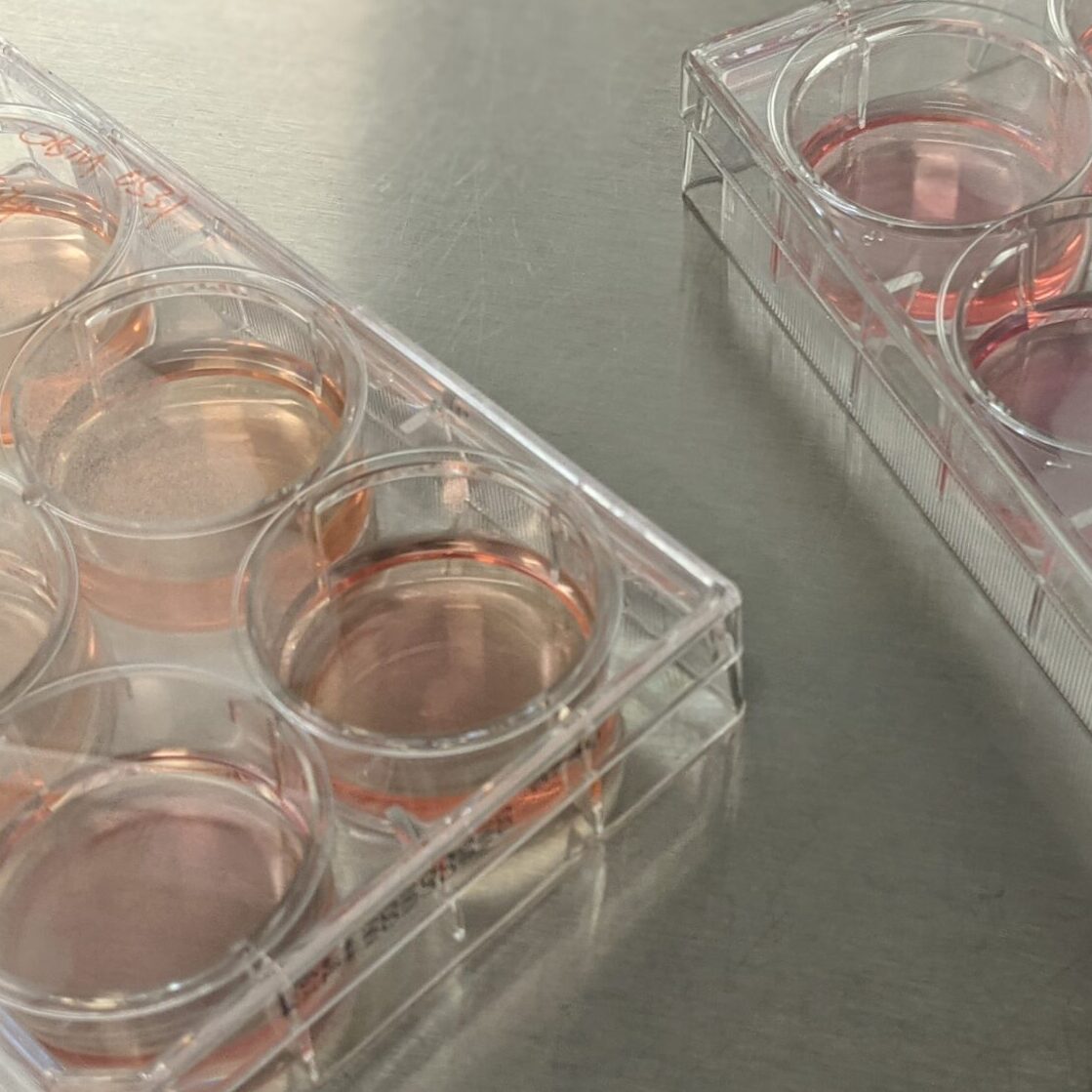
The Lentiviral Core Platform (LCP) produces high quality lentiviral particles at low cost for Manitoba researchers and houses 4 lentiviral vector shRNA libraries and two human ORFeome collections. The core stocks 293T cells, packaging and envelope plasmids for lentiviral production, control lentiviral vector plasmids, and an EGFP-expressing lentiviral vector. Services offered include custom-made viral preparations and EGFP-reporter and control viral preparations.
The LCP is located in the Apotex Centre, 4th floor and is maintained and operated by a full-time technician under the supervision of Drs. Sam Kung and Juyong Xie.